 |
 |
|
Field Emission SEM
The scanning electron microscopy is a versatile, non-destructive technique that reveals detailed information about the morphology and the composition of natural and manufactured materials. The SUPRA™ 40 (ZEISS) has been developed as an extremely versatile FESEM capable of delivering high quality imaging solutions for the many demanding applications in the field of nano technology. It offers high stability and a large (130 mm) fully motorised eucentric versatile specimen holder. Specially worth mentioning is that the GEMINI FESEM column utilised on the SUPRA™ 40 has an extremely low magnetic field outside the objective lens enabling investigation of magnetic materials and devices. A field-emission cathode in the electron gun provides narrower probing beams at low as well as high electron energy, resulting in both improved spatial resolution and minimized sample charging and damage. Performances:
Energy Dispersive X-rays detector In addition to the standard electron microscope detectors, The SUPRA™ 40 microscope is equipped with an Energy Dispersive X-rays detector EDX (EDAX) which is a major breakthrough in Si(Li) detecting technology. It exhibits unique features such as low-profile dewar and special cold finger design which allows complete cool-down from a warm state in less than one hour. This detector is designed to be left at room temperature and is completely unaffected by repeated cycling and it includes all circuitry necessary for powering down the detector and cycling it back to room temperature. In addition to the rapid warm-cold cyclability, this detector exhibits reduced L-N2 consuption and it is configurable for light element analysis down to beryllium.
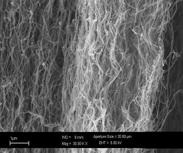
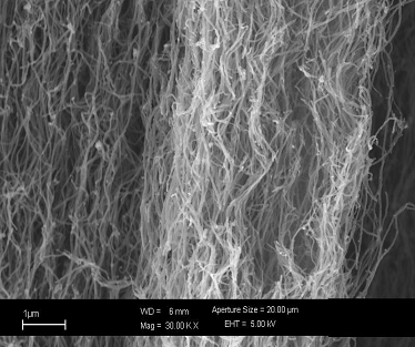
The FESEM can be classified as a high vacuum instrument (less than 1x10⁻⁷ Pa in the gun zone). The vacuum allows electron movement along the column without scattering and helps prevent discharges inside the gun zone. The function of the electron gun is to provide a large and stable current in a small beam. There are two classes of emission source: thermionic emitter and field emitter. Emitter type is the main difference between the Scanning Electron Microscope (SEM) and the Field Emission Scanning Electron Microscope (FESEM). Thermionic Emitters use electrical current to heat up a filament; the two most common materials used for filaments are Tungsten (W) and Lanthanun Hexaboride (LaB6). When the heat is enough to overcome the work function of the filament material, the electrons can escape from the material itself. Thermionic sources have relative low brightness, evaporation of cathode material and thermal drift during operation. Field Emission is one way of generating electrons that avoids these problems. A Field Emission Gun (FEG); also called a cold cathode field emitter, does not heat the filament. The emission is reached by placing the filament in a huge electrical potential gradient. The FEG is usually a wire of Tungsten (W) fashioned into a sharp point. The significance of the small tip radius (~ 100 nm) is that an electric field can be concentrated to an extreme level, becoming so big that the work function of the material is lowered and electrons can leave the cathode. FESEM uses Field Emission Gun producing a cleaner image, less electrostatic distortions and spatial resolution < 2nm (that means 3 or 6 times better than SEM). The FESEM has two anodes for electrostatic focusing. A voltage (0 ~ 5 KV) between the field emission tip and the first anode, called the extraction voltage, controls the current emission (1 ~ 20 micro-A). A voltage (1 ~ 30KV), called the accelerating voltage, between the cathode and the second anode increases the beam energy and determines the velocity at which the electrons move into the column. This voltage combined with the beam diameter determines the resolution (capacity to resolves two closely spaced point as two separates entities). As voltage increases, better point-to-point resolution can be reached. To resolve a feature on the specimen surface, the beam diameter must be smaller than the feature (still containing high current density). Therefore is necessary to condense the electron beam. To assist in the demagnification of the beam, electromagnetic lenses are employed. The table bellow shows the cross over diameter, final spot diameter and demagnification for thermoionic and field emitter. Since the cross over diameter in the Field Emission Source is smaller, a lower level of the beam condensation is necessary to have a probe useful for image processing. This makes the FESEM the highest resolution instrument. Variable apertures are used to refine the beam. Small objective aperture sizes will produce better resolution, good depth of field and minimal charging. It is the responsibility of the user to choose the correct aperture size. Depth of Field is important in routine microscopy and represents the ability to maintain the focus even with large changes in specimen topography. Long working distance (the distance from the final objective lens to the specimen surface) and small aperture yields images that appear in focus over a large change in Z-axis. The specimen and the electron beam interact in both elastic and inelastic fashion giving different types of signals. Elastic scattering events are those that do not affect the kinetic energy of the electron even when it's trajectory had been affected. Inelastic scattering events are a result of the energy transfer from the electron beam to the atoms in the specimen, as result the electrons have energy loss with small trajectory deviation. Each of these signals gets specific information about topography, crystallography, surface characteristics, specimen composition. CNT SEM Image
 View in High res
 View in High res
|
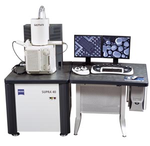 A brief description of the ZEISS SUPRA™ 40 Field Emission Scanning Electron Microscope (FESEM)
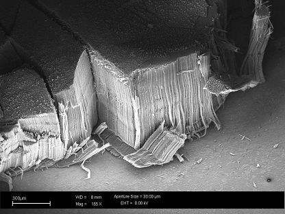