Nano-biosensing for Healthcare
With the achievements of nanotechnology, biosensing starts to take advantage of a wide variety of nanoscale materials and phenomena. Nanobiosensing opens up novel concepts in basic research and new tools for ultrasensitive biodetection in clinical and industrial applications. Extremely low detection limits, even reaching the single biomolecule level, have been demonstrated. Nanobiosensors not only enhance the detection capabilities, but also promise rapid, inexpensive, portable and label-free tools.
In our group, nanobiosensing activities are based on twomain approaches:
Nanomechanical sensing
Micro and nanomechanical resonators have been widely used in the last decades as highly sensitive mass detectors for biological and chemical applications. Since more than ten years, we are developing bioassays based on microcantilever resonator arrays, able to successfully detect minimum concentrations of target molecules such as tumor biomarkers, allergens, and carcinogenic small molecules, as well as single pathogenic bacteria.
More recently, new approaches are catalyzing the attention of the scientific community, such as Suspended Microchannel Resonators (SMRs) to minimize the damping associated with the fluidic viscous drag, 1D and 2D materials (CNT, graphene) to increase the mass sensitivity down to yoctogram range, and nanomechanical Mass Spectrometry (NEMS-MS) to identify mass and position of single particles.
In such a scenario, our current research (more details here) is focusing on:
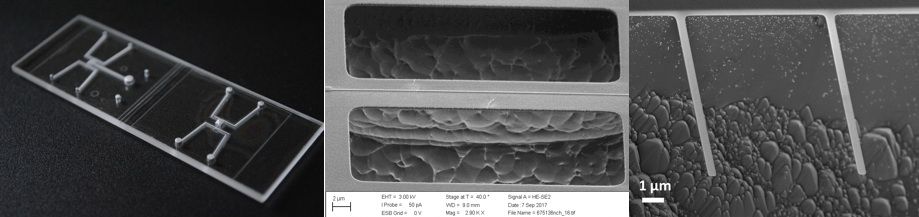
- monolithic and totally transparent Suspended Microchannel Resonators (SMRs)
- nanofluidic resonators towards single nanoparticle characterization in liquid
- nanomechanical resonators based on 1D and 2D nanostructures of inorganic and biological materials
Contact information
Carlo Ricciardi
+39 011 090 7398
carlo.ricciardi@polito.it
Publications
- “Development of a microcantilever-based immunosensing method for mycotoxin detection”, C. Ricciardi, R. Castagna, I. Ferrante, F. Frascella, S.L. Marasso, A. Ricci, G. Canavese, A. Lorè, A. Prelle, M.L. Gullino, D. Spadaro. Biosensors and Bioelectronics 40 (2013) 233–239.
- “Immunodetection of 17β-estradiol in serum at ppt level by microcantilever resonators”,C. Ricciardi, I. Ferrante, R. Castagna, F. Frascella, S.L. Marasso, K. Santoro, M. Gili, D. Pitardi, M. Pezzolato, E. Bozzetta. Biosensors and Bioelectronics 40 (2013) 407–411.
- “Functionalization protocols of silicon micro/nano-mechanical biosensors”, F. Frascella, C. Ricciardi. Nanomaterial Interfaces in Biology Nanomaterial Interfaces in Biology. Springer (2013) pp. 109-115.
- “Microcantilver-based DNA hybridization sensors for Salmonella identification”, R. Patti, M.T. Bottero, A. Dalmasso, A. Grassi, I. Ferrante, K. Santoro, N. Ciprianetti, C. Ricciardi. Italian Journal of Food Safety Vol. 1 (2012) 17-19, ISSN: 2239-7132.
- “Online Portable Microcantilever Biosensors for Salmonella enterica Serotype Enteritidis Detection” C. Ricciardi, G. Canavese, R. Castagna, G. Digregorio, I. Ferrante, S.L. Marasso, A. Ricci, V. Alessandria, K. Rantsiou, L.S. Cocolin. Food Bioprocess Technol 3 (2010) 956–960.
- Development of microcantilever-based biosensor array to detect Angiopoietin-1, a marker of tumor angiogenesis”, C. Ricciardi, S. Fiorilli, S. Bianco, G. Canavese, R. Castagna, I. Ferrante, G. Digregorio, S.L. Marasso, L. Napione, and F. Bussolino. Biosensors and Bioelectronics, 25 (2010) 1193-1198.
- “ Integration of microfluidic and cantilever technology for biosensing application in liquid environment” C. Ricciardi, G. Canavese, R. Castagna, I. Ferrante, A. Ricci, S. L. Marasso, L. Napione, F. Bussolino, Biosens. Bioelect., 26 (2010) 1565-1570
Plasmonic nanostructures for SERS biodetection
Surface Enhanced Raman Scattering (SERS) is a sensitive technique allowing vibrational spectra from individual molecules to be measured. Among single-molecule spectroscopies, it provides much more detailed information as compared to the broad fluorescence spectra. Actually, due to the almost unstructured spectra, fluorescence does not provide detailed molecular information, and photobleaching effects often inhibit single molecule analysis. Raman spectroscopy provides highly resolved vibrational information and although the molecular Raman cross sections are much smaller than the fluorescence ones, the SERS mechanism can enhance the Raman efficiency making it competitive in terms of signal intensity.
In the framework of this research activity, metal-dielectric nanostructures consisting of Ag nanoparticles are synthesized within a mesoporous silicon matrix (Ag/pSi) on large area by dip coating and ink-jet printing. These substrates are exploited for detection of biological assays approaching single molecule detection by surface-enhanced resonance Raman scattering (SERRS). The nanostructures morphology is controlled yielding plasmonic resonances in the visible-near-infrared range. Tuning the particle plasmonic resonance close to the molecule electronic resonance, we have demonstrated Raman enhancements larger than 10^10.
As representative examples, these efficient SERS-active substrates were successfully used for the detection of short peptides and miRNA, consisting of small non-coding single-stranded sequences that are of great relevance in gene regulation affecting process such as cell proliferation, cell death and oncogenesis. The recognized roles of these sequences suggest that some miRNA or pattern of miRNA can be used as biomarker for early cancer diagnosis. In particular, we optimized a protocol for the thiolated cDNA oligonucletides immobilization on the silver nanoparticles. The successful binding of -SH terminated cDNA on Ag nanoparticles was checked by SERS measurements and confirmed by ELISA analysis performed on flat Ag silicon substrates functionalized with the same protocols. Promising results were observed in tests concerning with cDNA-miRNA hybridization using blocking agents/spacers in a cDNA co-immobilization protocol. The Raman fingerprint of the cDNA-miRNA complex showed selectivity and reproducibility as required by most of the SERS applications on biological assays.

SERS spectra of the CSFNIT peptide chemisorbed on functionalized Ag/pSi, compared with the Raman spectra of the pure amino acids constituting the sequence.

SERS spectra concerning with the detection of cDNA-miRNA (5’[Cy5]AGCUACAUCUGGCUACUGGGU3’) hybridization on Ag/pSi
Contact information
Fabrizio Giorgis
Tel. +39 011 090 7354
Fabrizio.giorgis@polito.it
Publications
- “Silver Nanoparticles on Porous Silicon: Approaching Single Molecule Detection in Resonant SERS Regime”, Alessandro Virga, Paola Rivolo, Francesca Frascella, Angelo Angelini, Emiliano Descrovi, Francesco Geobaldo, Fabrizio Giorgis. Journal of Physical Chemistry C 117 (2013) 20139-20145.
- “Ag/pSi SERS platforms as biosensors for oligonucleotides/miRNA detection”, A.Virga, A. Chiadò, S. Ricciardi, F. Frascella, C. Novara, P. Rivolo, F. Geobaldo, F. Giorgis. Porous Semiconductors - Science and Technology– PSST2014 Proceedings p. 224.
- “SERS active Ag nanoparticles in mesoporous silicon: detection of organic molecules and peptide-antibody assays”, A. Virga, P. Rivolo, E. Descrovi, A. Chiolerio, G. Digregorio, F. Frascella, M. Soster, F. Bussolino, S. Marchiò, F. Geobaldo, F. Giorgis. Journal of Raman Spectroscopy 43 (2012) 730-736.
- “Direct patterning of silver particles on porous silicon by inkjet printing of a silver salt via in-situ reduction”, A. Chiolerio, A. Virga, P. Pandolfi, P. Martino, P. Rivolo, F. Geobaldo, F. Giorgis. Nanoscale Research Letters 7 (2012) 502-507.
- “Metal-dielectric nanostructures for amplified Raman and fluorescence spectroscopy”, Alessandro Virga, Rossana Gazia, Luca Pallavidino, Pietro Mandracci, Emiliano Descrovi, Angelica Chiodoni, Francesco Geobaldo, Fabrizio Giorgis. Physica Status Solidi C 7 (2010) 1196-1199.
- “Porous Silicon as efficient Surface-Enhanced Raman Scattering (SERS) Substrate”, F. Giorgis, E. Descrovi, A. Chiodoni, E. Froner, M. Scarpa, A. Venturello, F. Geobaldo. Applied Surface Science 254 (2008) 7494-7497.